Numerical Simulation and Thermal Analysis of Tumor in the Human Body
INTRODUCTION: Abnormalities in local body surface temperature have been recognized as a sign of disease for centuries, much before humans knew about the cause of ailments or of pain [1]. The idea of this work is to use numerical simulation tools to predict the location, size and metabolism of tumor embedded in any outer body organ of human. Idealized thermal data of an organ, modeled either as a solid rectangle, cylinder or hemisphere respecting to its physical structure, which encompasses a hyperactive region, modeled spherical heat source, has been obtained using finite element method by solving the steady-state Pennes’ bioheat equation with non-linear boundary conditions [2].
USE OF THE COMSOL MULTIPHYSICS® SOFTWARE: Numerical simulation using the COMSOL Multiphysics® software was performed to solve the problem stated in INTRODUCTION. Developed 3-D finite element models for the three different physical cases to study the bio-thermal properties of the tissues with tumor (hot nodule embedded in healthy organ) are shown in Figure 1. Mesh sensitivity analysis were performed to determine the optimum sizes in order to obtain a good resolution in the simulated patterns, and to minimize the computational time. In addition, nodes along the surface area constrained in the normal-translational direction for all three geometries. All other nodes are unconstrained in all directions. A constant temperature of 293.15 K was applied at the entire section as an initial condition.
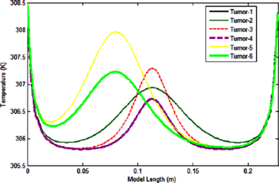
RESULTS: The simulated temperature patterns at skin surface and tissue interior of rectangular box tissues are presented in the following Figure 2. In Figure 2 at left, the 1-D temperature distribution along the line parallel to the xy- plane and directly above the tumor center is presented, which shows that a difference in temperature is 0.15o between the region that is directly above the tumor and regions those are away from it; Figure 2, right, presents its 2-D temperature evolution over the skin surface that bears the sign over which the temperature grows; and also a three level isothermal boundaries having around 0.1 C separation is shown in the isothermal contour plot in the Figure 2 at right.
CONCLUSION: A methodology was developed for the estimation of thermophysical or geometrical parameters of tumor region using the temperature profile on the skin surface. The problem was solved using COMSOL Multiphysics® software to model tissues with different geometry. According to the results, the methodology can help to locate tumor region on any external body part which could be useful and important to study tumor evolution after a treatment procedure.
Download
- mohammadi_poster.pdf - 0.49MB
- mohammadi_abstract.pdf - 0.26MB