Acoustic Propagation Model in 3D of Dolphin Echolocation Formation Inside Head Tissues
Introduction The echolocation signals of dolphins consist of forward-projected sound pulses that reflect off objects and are used to probe underwater surroundings. Despite rigorous studies of dolphin echolocation, there remain questions about how echolocation sounds are produced and shaped into an intricate forward projected beam. Most previous acoustic models of echolocation sound propagation through dolphin head tissues were conducted in 2D and were based on computed tomography (CT) scans of dolphin carcasses. Modelling dolphin echolocation in 2D is insufficient as no 2D plane of the animal can accurately approximate the asymmetric 3D anatomy. One previous study used in vivo CT scans with 3D modeling, however without fully exploiting the potential of using subject-specific 3D modelling for instance when it comes to implementing detailed tissue-specific acoustic properties. In this study, we present a full bandwidth, 3D finite element model (FEM) of bottlenose dolphin echolocation signal propagation in which each mesh node has its own material properties based on in vivo CT scans.
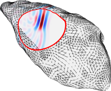
Methods Dolphin echolocation sounds are produced in the nasal passages and travel through the “melon”, a structure located in the forehead that acts as an acoustic collimating lens. The modelled shape of the melon was generated through manual segmentation based on the Hounsfield units (HU) of in vivo CT scans of an adult bottlenose dolphin. The density and sound velocity of each mesh node of the melon were derived using an empirically approximated relationship between HU, density, and sound velocity and implemented via an interpolation function describing the CT based HU values in COMSOL Multiphysics® (Fig. 1). The melon was placed inside a block of modelled water surrounded by an acoustically absorbing layer. The sound source was modelled as a vibrating half sphere, located at the site of echolocation signal generation (upper left of the melon in Fig 1), generating an acoustic pressure wave that propagates into the melon. The source was modelled as a transient time signal containing the full frequency bandwidth of bottlenose dolphin echolocation signals (Fig. 2). The final model was solved in time domain using the discontinuous Galerkin method and the Acoustics Module in COMSOL®.
Results Our results show how the properties of the melon concentrate the acoustic energy into a forward projected beam, Fig. 3(a). Conversely, modelling the melon as a homogenous medium with water-like properties resulted in a uniform distribution of the acoustic energy Fig. 3(b), with no wavefront collimation.
Discussion We produced the first fully 3D model of the dolphin melon using in vivo CT scans with full bandwidth. Our results show that heterogeneous density and acoustic properties are crucial to replicating the collimating effect of the melon, as well as modelling this in 3D as opposed to 2D. This is an important step towards a thorough understanding of how echolocation works in dolphins. Nasal air pockets and bone structures inside the dolphin head tissues are believed to play a fundamental role in beam formation and are likely involved in beam steering and beamwidth control, both of which have been observed in dolphins. Future work will focus on including such features in a fully 3D model.
Download
- Starkhammar_4561_poster.pdf - 3.03MB